Label the Structures of the Finger and Fingernail
It lies under the cuticle. The nail plate should NOT be confused with the nail bed.
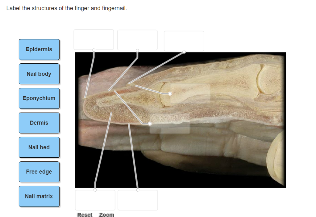
Solved Label The Structures Of The Finger And Fingernail Chegg Com
Dorsal surface back of the hand containing the fingernails at the tips.
. Start studying Labeling the Structure of a Nail. The cuticle of the fingernail is also called the eponychium. The ends of the toes and fingers are protected by the nails which are formed from nail roots present under the skin folds at the sides and base of the nail.
The finger bones are known as phalanges singular phalanx. The nail bed is the skin under the nail. There is a printable worksheet available for download here so you can take the quiz with pen and paper.
This is attached to the nail bed and appears as white. Visible Part of the matrix that extends from underneath the living skin. The thickness of your nails is determined by the size of your matrix.
Keratin is the main component of a. Fingernails and toenails are made of a tough protein called keratin as are animals hooves and horns. The average person has 50 layers of keratin cells that make up the nail plate.
We also discuss some of the f. You need to get 100 to score the 11 points available. An extra dot has been applied to this gameThat is why score were reset.
Free edge of nail. Dead colorless tissue attached to the natural nail plate. This illustration will help you know all the proper names and locations of the parts that make up a single nail tip.
Epidermis Nail body Eponychium Dermis Nail. The nail grows longer as the new cells are formed. The cuticle is situated between the skin of the finger and the nail plate fusing these structures together and providing a waterproof barrier.
Learn vocabulary terms and more with flashcards games and other study tools. The function of the free edge is to protect the fingertip and the hyponychium. Palmar surface front of the hand continuous with the palms of the hand.
Identify label the parts of the nail structure and give the function of each part. A specialized form of epidermis that is found over the base of the nails of the fingers. Along with hair they are an appendage of the skin.
Learn to speak fluent nail by knowing your anatomy. We cover the different parts of nails and how nails grow. Experts are tested by Chegg as specialists in their subject area.
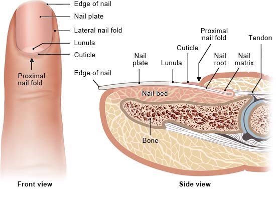
The skin on both sides of the nail plate. Is the white crescent-shaped area of a finger. The nail plate is the hard nail itself.
A fingernail consists of several parts including the nail plate the visible part of the nail the nail bed the skin beneath the nail plate the cuticle the tissue that overlaps the plate and rims the base of the nail the nail folds the skin folds that frame and support the nail on three sides the lunula the. The matrix is the tissue from which the nails grow. Thin layer of tissue that attaches the nail plate and the nail bed.
We review their content and use your feedback to keep the quality high. Like a lunar moon Cuticle. This is a part of the epidermis under the free edge of the nail plate.
Skin folds anchor the nails to the fingers. Our solutions are written by Chegg experts so. In this video we discuss the structure of fingernails and toenails.
Human anatomy Basic nail anatomy The nail consists of the nail plate the nail matrix and the nail bed below it and the grooves surrounding it1 Parts of the nail The matrix synonyms2 matrix unguis keratogenous. Access Student Worksheets for Visual Anatomy and Physiology 1st Edition Chapter 5 Problem 2SR2 solution now. This is the part of the finger underneath the nail plate.
Your Skills Rank. Draw a fingernail in your notebook. The nail plate leaves the end of the finger and forms a projection that is called the free edge.
This is an onlin equiz about the anatomy of the finger top. Label the structures of the finger and fingernail. A fingernail is produced by living skin cells in the finger.
This is the skin that frames each of your nails on three sides. Cuticles are the tissues along the sides and the base of nails. This is an online quiz called Nail Structure.
This is the part we file and shape. It is whitish half-moon shaped at the base of the nail. There are 14.
The Free Edge. The lunula is the visible half-moon at the base of the nail. 5th finger little finger pinky digitus minimus manus There are two surfaces of the fingers.
Your nail bed is the skin beneath the nail plate. Learning Task 6. These are the grooves on the skin at the sides of the free edge and the nail follows them as a guideline when it grows.
Composed of hardened flat translucent non-living keratin nail cells that form a solid protective layer over the underlying soft tissue.

Pin On Faith
Structure Of The Nails Informedhealth Org

Pin By Adri Roelofse On Coding Anatomy Physiology Types Of Nails Nails School Nails


Comments
Post a Comment